1. Axons shear due to strain, especially at the junction of white matter and grey matter.
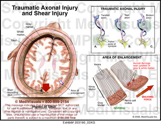
2. This shearing is vvisible microscopically, but not through neuroradiography (e.g., CT/MRI)
Axonal spheroids in shear-type NAHI. (a) High-power photomicrograph (original magnification ×400; hematoxylin-eosin stain) of the medullary corticospinal tract from a 3-month-old infant reveals axonal injury as axonal enlargements or spheroids (arrows) (ref)
High-power photomicrograph (original magnification, ×400; βAPP with hematoxylin stain) of a pontine corticospinal tract from a 2-year-old child clearly shows axonal spheroids (arrows). THese are the "retraction balls" we spoke of... (ref)
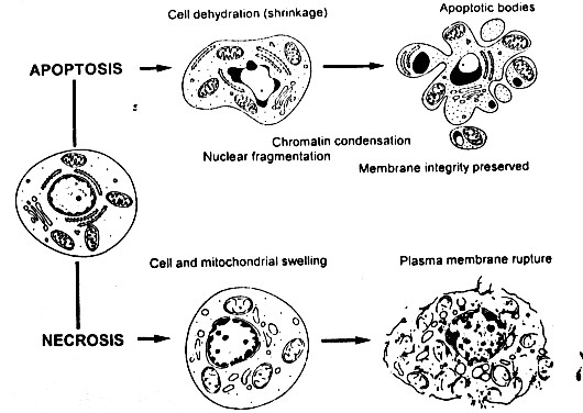
3. Cells can undergo apoptosis or necrosis
4. Axons can become "swollen" or damaged
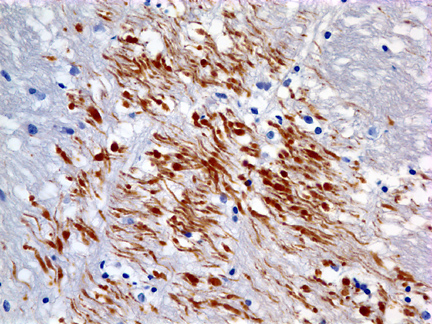
brown stain=swollen axons
5. Damaged axons lead to influx of "normally excluded extracellular ions", such as calcium.
6. The presence of these metabolites can be measured using MRI spectroscopy
Figure reveals MRS values for a patient with very severe head trauma at two separate time points (Day 9 and Day 25). Note diminished NAA concentrations and elevated Cho indicating significant brain damage. In addition, lactate is the highest peak at Day 9, with reductions by Day 25. Lactate elevations coincided with reductions in ICP, which may be indicative of an ischemic process. (ref)
7. Changes in glucose metabolism follow:
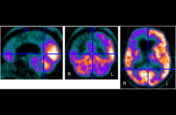
Following stroke
Following coma ref)
8. This leads to oxidative stress
(APP=amyloid precursor protein; Aβ=β-amyloid; bcl-2=B-cell lymphoma 2)
9. Changes in downstream axonal communications can occur: DTI