 A widely accepted theory of brain functioning was proposed by Luria. In his book, The Working Brain, he outlined "three principal functional units of the brain whose participation is necessary for any type of mental activity" (Luria, 1974, p. 43). These units are responsible for regulating cortical tone or waking, for obtaining, processing, and storing information arriving from the outside world, and for programming, regulating, and verifying mental activity. Luria (1974, p. 43) also proposed that each of these units is "heirarchical in structure and consists of at least three cortical zones built one above the other". A primary "projection" area receives impulses from or sends impulses to the periphery. A secondary "projection-association" area processes incoming information and programs information for projection to efferent pathways. The tertiary "zones of overlapping" area is last to develop and is responsible for complex forms of mental activity which requires the integrated participation of many cortical structures. These units and zones, when functioning properly, work together to regulate all our behaviors, from waking and sleeping, to hearing and seeing, and thinking and problem solving.
The unit for regulating tone, waking, and mental states lies below the cerebral cortex and is commonly known as the reticular activating system (RAS). This unit has a dual relationship with the cortex, in that the RAS both influences the tone of the cortex, and also experiences a regulatory influence (Luria, 1974). Through the cells of this unit, excitation spreads gradually, changing their levels little by little. This is in direct contrast to the cells in the cortex which send single impulses along their long axons operating on an "all or nothing" law which states that the cell will store up energy until there is enough to depolarize and send an impulse. Instead, the cells in the RAS form a "nerve net" in which the bodies of cells are connected by relatively short axons. Excitation which gradually spreads through the RAS will ultimately excite the cortex and, according to Luria (1974), modulate the whole state of the nervous system.
The fibers of the RAS form pathways which are both ascending and descending and allow the RAS to have a reciprical relationship with the cerebral cortex. The ascending pathways of the RAS synapse with higher level structures in the nervous system such as the thalamus, caudate body, and ultimately the neocortex. This ascending pathway gradually spreads excitation upwards to "activate the cortex and regulate the state of its activity" (Luria, 1974, p. 46). The descending pathways of the RAS begin in the higher level structures starting in the neocortex and run to lower structures synapsing ultimately in the brain stem. This descending pathway allows the higher structures to "subordinate these lower structures to the control of programmes (axons) arising in the cortex and requiring modification and modulation of the state of waking for their performance" (Luria, 1974, p. 46).
So, the first functional unit not only changes the tone of the cortex, but is also under control of the cortex, allowing the RAS to help the nervous system to respond and adapt to perceived changes in the environment. Thus, the heirarchical organization of the nervous system proposed by Luria assumes that higher structures in the system are dependent upon the lower structures for activation as well as regulation and maintenance of the activation. This activation is derived from three sources: metabolic processes, the arrival of stimuli from the outside world activating an "orienting response", and internal plans or goals which evoke the activation of neurons leading to the attainment of the goal. Any disruption in the ascending or descending RAS pathways, or damage to the processes and structures which activate this functional unit, will result in an insufficient state of waking or cortical tone, which in turn results in an organism which can not sufficiently interact with its environment.
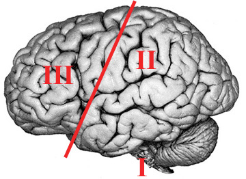
The second functional unit is primarily responsible for the reception, analysis, and storage of information. This unit occupies the posterior region of the neocortex, including the occipital, temporal, and parietal lobes, and plays a vital part in bringing visual, auditory, gustatory, olefactory, vestibular, and general sensory information into the cortex (Luria, 1974). The structures comprising this unit consist of isolated groups of neurons in parts of the cortex which receive impulses and relay impulses to other neurons. The primary zones of these structural units employ modality-specific groups of neurons to receive impulses from the sensory organs, while the secondary zones of these structures surround the primary zones with associative neurons which enable incoming excitation to be conveyed to the tertiary zones. These tertiary zones, or "zones of overlapping", are responsible for integrating and organizing the excitation arriving from the different sensory structures, and converting the successive stimuli into simultaneously processed groups (Luria, 1974). Any damage to the structures forming the second functional unit can result in decreased efferent impulses to orient the first functional unit, or incomplete information being transmitted to the third functional unit.
The third functional unit is responsible for programming, regulating, and verifying conscious activity. Forming plans and intentions, regulating behaviors, monitoring progress towards goals, and correcting mistakes are all activities associated with this third functional unit; located in the regions anterior to the precentral gyrus (Luria, 1974). Neural activity passes through this unit to the primary motor cortex where impulses are transmitted into motor routines and speech patterns. These impulses are projected first to the secondary zone of the third functional unit, incorporated in the premotor areas of the frontal region. It is within this premotor area that neural activity is transmitted to systematically organized movements (such as grasping movements of the hands, turning the head and eyes, or forming words and sentences instead of individualized twitches of muscles) before passing through the structures of the primary motor cortex to the periphery (Luria, 1974). This prefrontal area also connects with lower levels of the brain and is instrumental in modulating the activities in these lower levels. Afferent impulses from all areas of the brain are synthesized in the prefrontal structures and organized for efferent projection, thus inhibiting or activating behaviors controlled by the afferent areas. Damage to the third functional unit can alter this regulatory control by impairing the ability of the prefrontal area to synthesize and organize these impulses, resulting in a dissociation between the afferent impulses and the behaviors which arise from the efferent activity. In addition, damage to the prefrontal area can alter the reciprocal relationship between cortex and the RAS, so that the brain may not be sufficiently aroused for complex behaviors requiring sustained attention.
"Each form of conscious activity is always a complex functional system and takes place through the combined working of all three brain units, each making its own contribution" (Luria, 1974, p.99). In order to perform a voluntary movement, according to Luria (1974), the systems of the first unit provide the muscle tone, the systems of the second unit provide afferent feedback as to the status of the movement, while the third unit regulates the movement by synthesizing the neural activity and coordinating and adjusting the movement toward the goal. Similar scripts can be outlined for virtually any act of perception, verbalization, audition, motion, etc. However, when this complex functional system is damaged by injury to any or all of the units, the cohesion of the system is disrupted, resulting in a system which functions in a manner markedly different than before the disruption.
Mechanism of Injury
Lobes of the Brain
Click here to return to the Neuropsychology Course page.